
Cholesteatoma is a distinct disease that affects the middle ear. It is characterized by the growth of a non-cancerous tissue in the middle ear. Though non-cancerous, the tissue can grow in size and destroy the neighboring tissues inside the ear. This can lead to an array of cholesteatoma symptoms. The growth of this cyst can damage structures of the ear, including the tiny bones and other structures responsible for balance and hearing.
What Are the Cholesteatoma Symptoms?
At the beginning of the infection, its symptoms are mild. However, as the cyst continues to grow, the symptoms become more severe. Among the initial symptoms of this condition is a smelly discharge from the ear. With time, pressure will start to build up in the ear causing discomfort. This may be accompanied by an aching pain either behind or in the ear. This increased pressure within the ear can also lead to deafness. If left unattended to, the cyst will enlarge and affect the face as well, causing facial muscles to either weaken or paralyze. At this point of cyst growth, the patient may also start feeling dizzy.
Other possible cholesteatoma symptoms may also include:
- Infection in the ear, leading to a smelly ear
- Temporary or permanent deafness
- Vertigo, a feeling that the environment around you is spinning
- Tinnitus, a perception that sound is coming from inside the body
- Weakening of facial muscles on half of the face
- Labyrinths
- Brain abscess
- Continuing ear drainage
- Meningitis
- The cyst may spread to the brain
What Causes Cholesteatoma?
Various theories exist regarding the formation of cholesteatoma. The majority of evidence collected to date seem to suggest that this condition is caused by malfunctioning of the Eustachian tube. When the eustachian tube fails to open and equalize the pressure in the middle ear, a negative pressure develops behind the eardrum. This in turn retracts the eardrum to form a pocket-like structure. As the pocket-like structure deepens, the skin gets trapped in between the ear as a sac or cyst and dead skin cells slough off. Unfortunately, this will also be happening to the cholesteatoma sac. Dead cells continue to slough off, thus expanding the sac and forming a cholesteatoma. In some instances, skin will grow around a margin of perforation in the middle ear.
Is It Common?
The condition is very rare, with statistics suggesting that only one in every 10,000 people suffers from this condition. In every 1,000 patients referred to an ear, nose and throat (ENT) clinics with a hearing problem, only one is likely to suffer from cholesteatoma. Most of the cholesteatoma cases are of the acquired type.
How Is CholesteatomaDiagonosed?
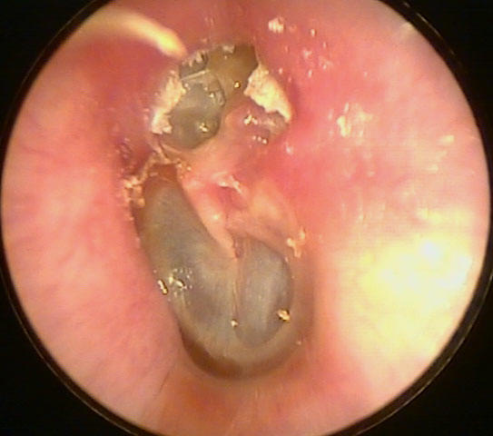
The method used to diagnose this condition mainly depends on the symptoms. For instance, if the patient complains of pressure, pain, hearing loss and drainage in the ear, the doctor will use an otoscope to examine the interior of the ear. With the device, the doctor can see any mass of blood vessels or deposit of skin cells, which are indicators of a growing cyst. Additionally, the doctor will also be able to notice any hole in the eardrum caused by the suction force that led to the development of the cyst.
If the symptoms are mild, such as facial muscle weakness and dizziness, the doctor will need to use a computed tomography (CT) scan. This technology captures a cross section image of the inner side of the ear. The image gives the doctor a clear view of the inside of the skull and the ear and enabling him or her to determine the cause of your symptoms.
How Will Cholesteatoma Be Treated?
Preliminary treatment is aimed at stopping the growth of the cyst and stop drainage within the ear and may comprise of antibiotics, ear drops and careful cleaning of the ear.
If the cholesteatoma is complicated, a surgical treatment is required to prevent serious complications. To use this form of treatment, CT scans, balance and hearing tests of the mastoid may be required. Such tests are meant to estimate the hearing capacity of the ear and determine the level of damage caused by the cyst. In most cases, this surgical procedure is conducted under an anesthesia to remove the cholesteotoma and result in a dry ear. After this initial surgery, a second one, aimed at ensuring that the cholesteatoma is completely gone and reconstructing the damaged structures of the ear, may be necessary.
If the condition is severe, a reconstruction surgery may not be possible. Additionally, procedures to regulate dizziness or repair facial muscles are not always essential. The reconstructive surgery is normally done 6 to 12 months after the initial operation. The aim of the reconstructive surgery is to restore hearing and inspect middle ear to ensure that the cyst is completely removed.
A surgical procedure to remove the cyst is performed as an out-patient procedure, with most of the patients being required to stay overnight. In rare cases, prolonged hospitalization may be necessary for an antibiotic treatment. This is particularly when the condition is serious. To undergo this procedure, you will require one to two weeks time off from work. After the procedure, you will need to visit your doctor regularly to check for recurrence and evaluate the outcome. For cases requiring creation of a mastoidectomy cavity, you will be required to visit the doctor once every few months to have the mastoid cavity cleaned in a bid to prevent infections.