
The small intestine villi are the very small finger type projections protruding from the epithelial lining in the intestinal wall. The villi are about 0.5 to 1.6 mm in length. The villi contract and expand which allows for the increase in surface, thus enhancing nutrients absorption.
The Function of Small Intestine Villi
The purpose of the villi is to help to digest the nutrients in the foods that are eaten. It helps the nutrients to be passed through and effectively used in the body as required. The villi are also connected to blood vessels and the blood that circulates then carries the nutrients away to be eliminated when they are not being used by the body. All of this process is going on automatically anytime a person eats. It's part of the digestive system.
How It Works
As a part of the intestines in the gastrointestinal tract, this is where a major portion of the foods are digested. The main reason for the small intestines is to absorb nutrients and the minerals that are found in the foods we eat and to prepare them to properly fuel the body. Without proper absorption, the foods wouldn't be able to provide the nutrients that the body requires to function properly.
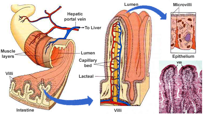 As the nutrients pass from the blood vessels via the walls of the intestines, they undergo a process called diffusion. Here the inner walls or the mucosa as they are called, are lined with tissue called columnar epithelial tissue. This tissue is a permanent feature and it allows the intestines to contract and distend. They also have finger type projectiles which are called microvilli (villi is Latin for "shaggy hair"). The small intestine villi and the microvilli are there to help increase the surface area that absorbs the nutrients and to allow for more absorption thus improving the function of the body.
Each and every one of the small intestine villi has their own unique network of capillaries. These capillaries also have fine lymph vessels which are called lacteals. The lacteals reside close to the surface. Here, nutrients are transported from the lumen of the intestines. They move and combine with amino acids and with carbohydrates (or carbs as they are commonly called) and with the lacteals or lipids. Everything that is absorbed is moved through the blood vessels into the various organs in the body where they combine to create the complex proteins that your body requires to survive. Any remaining foods are then passed into the large intestines where they are eventually eliminated through having a bowel movement.
Celiac Disease and Villi
If your intestines become inflamed, it can greatly affect your digestion and the absorption of the nutrients and your minerals. One of the most common culprits of this is celiac disease. Celiac disease is an immune reaction to the protein, gluten. Gluten is found in wheat, rye and barley.
Gluten sensitivity and celiac disease both can greatly impact the entire digestive process as well as the absorption of the nutrients that the body requires. Both are autoimmune conditions that can damage your small intestine villi and cause distress in absorption of the foods that are eaten.
Effects Caused
There are two major effects of the conditions: The first major effect is the damage and eventual destruction of the small intestine villi which reduce the amount of surface areas in the intestines. When these are damaged, the foods that are eaten will no longer properly digest and nourish the body.
Secondly, you won't be getting the nutrients that your body requires to function properly and thus you are at more risk for malnutrition because of vitamin and mineral deficiencies.
Over the course of time, patients may start to feel fatigued, suffer from anemia and even bone loss. The body will be at an increased risk for loss of weight and for seizures. Celiac disease can be readily tested for by a doctor and it is often something that the doctor will check for if the patient is consistently fatigued and has anemia.
Small Intestine Anatomy
 Measuring approximately 10 feet in length, the small intestine gets its name due to the fact that's it's a mere 1 inch in diameter, less than half the diameter of the large intestine. This convoluted tube of the digestive system will absorb approximately 90 percent of the nutrients in the foods that are eaten. These nutrients then pass through the body by blood that is circulating through the small intestine villi. They then move the nutrients out of the digestive tract and into system where they are used as required to properly nourish the body.
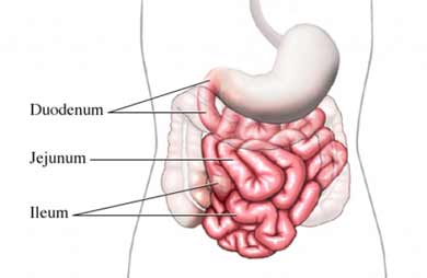
1. Duodenum
The first and the shortest portion of the small intestine is the duodenum. Partially digested foods pass through here via the stomach. The duodenum helps to chemically digest the chyme (partially digested foods) and prepares them for being absorbed into the smaller intestines. Without this preparation, the foods wouldn't be properly used by the body. To properly prepare the foods, chemicals from the pancreas, the liver, as well as the gall bladder are mixed in with the chyme to help the digestive process in the body.
2. Duodenojejunal Flexure
The portion of the duodenum and the jejunum join is called the duodenojejunal flexure. This formation follows the C shape as it's passed in front of the kidneys and into the upper three lumbar vertebrae and the duodenojejunal flexure ends where it joins the jejunum.
3. Jejunum
As the mid-section of the smaller intestines, the jejunum serves as a site for nutrients to be absorbed into the body. It measures approximately 3 feet and helps to improve nutrient absorption in the body. Without this step, the nutrients wouldn't be properly prepared for utilization.
4. Ileum
The final section of the small intestine is the ileum. The ileum empties into the larger intestines through the ileocecal sphincter. It's approximately six feet in length and it completes the absorption process of the nutrients.
5. Terminal Ileum
As the distal end of the smaller intestines, the terminal ileum is the portion of the smaller intestines that intersects together with the larger intestine. Containing the ileocecal sphincter, this is the smooth portion of muscle that controls how the chyme flows into the larger intestine. On the right side of the abdominal cavity, it is located near the umbilical and the hypogastric region of the abdominal cavity. Measuring in at approximately 1.25 to 1.5 inches long at the end, it intersects with the end of the ileum and ends at the ileocecal sphincter which is the band of muscle that is flowing into the chyme.
6. Ileocecal Valve
Also known as the Tulp's valve, it's located where the large and the small intestines unite. This valve restricts the flow of fluids to just one direction. It is circular in shape and contracts as required to help limit the reflux of the colonic contents. About 2 liters of liquid will pass through the ileocecal valve into the colon on a daily basis. The ileocecal valve absorbs both bile and Vitamin B 12 and is the only part of the body to absorb bile as well as Vitamin B12.